
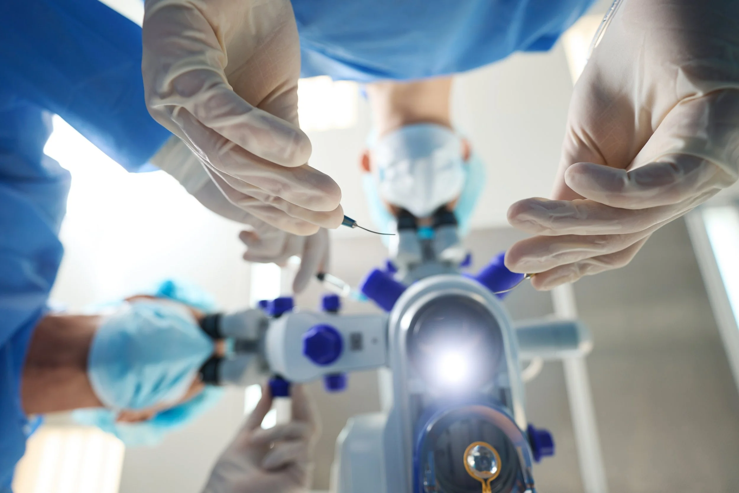
Cataract Surgery
Learn more about what a cataract is, your intraocular lens options, and frequently asked questions.
Understanding Cataracts
A cataract forms when the clear lens inside your eye becomes cloudy, which stops light from passing through as it should. This cloudiness can make your vision look blurred, dim, or hazy, much like looking through a fogged or frosted window. You might notice that colors seem duller, lights create more glare, and driving at night becomes harder.
Cataracts typically form slowly over time as part of the natural aging process, though they can also result from factors such as diabetes, prolonged sun exposure, eye injuries, or certain medications. In the early stages, vision changes may be subtle and can often be managed with stronger lighting, updated eyeglasses, or magnifying lenses.
As a cataract gets worse, the cloudiness can become so dense that it makes daily activities much harder. Reading, driving, or recognizing faces may become challenging. When this happens, doctors usually recommend cataract surgery.
Cataract surgery is one of the most commonly performed and safest procedures worldwide. During surgery, the cloudy natural lens is removed and replaced with a clear artificial lens, known as an intraocular lens (IOL), which restores sharper, brighter vision. Most patients experience significant improvement in visual clarity and quality of life shortly after surgery.
What Happens During Cataract Surgery
Cataract surgery is a precise microsurgical procedure and is performed on an outpatient basis, meaning patients go home the same day.
Before surgery begins, numbing eye drops are applied to ensure comfort, and a mild sedative may be given to help patients relax. Most individuals remain awake during the procedure but do not feel pain. While patients may notice bright lights or a sense of movement, they cannot see the surgical details.
The procedure involves several key steps:
Creating a tiny incision: Your surgeon makes a small opening at the edge of the cornea, usually less than 3 millimeters wide, using a special surgical tool or a laser.
Removing the cloudy lens: The front portion of the capsule surrounding the lens is carefully opened, and the cataract is broken apart using ultrasound energy. The lens fragments are then gently suctioned out.
Inserting the artificial lens: Once the natural lens has been removed, the surgeon places a foldable IOL through the same incision. The IOL unfolds inside the lens capsule and is precisely positioned to restore clear vision.
After the new lens is in place, the incision typically seals on its own without stitches. Your surgical team then examines the eye and applies a protective shield. Patients rest briefly in the recovery area before being discharged with detailed postoperative instructions and prescribed eye drops to prevent infection and promote healing.
Most patients notice clearer vision within a few days, although full visual recovery may take several weeks as the eye adjusts to the new lens.
Intraocular Lens Options
Frequently Asked Questions
-
Complications from cataract surgery are rare, but here is a video outlining some common side effects you may experience post surgery:
-
Each patient is different, so always consult with your surgeon. Here are a few general examples of things you can and cannot due immediately after surgery:
-
Healing times after cataract surgery can vary from person to person. How quickly vision improves depends on factors like the cataract’s severity, overall eye health, and the presence of other medical conditions. Most healthy patients start noticing better vision within a few days, with further progress occurring over the next three to four weeks.
-
Cataract surgery’s primary goal is to restore clear vision by taking out the cloudy lens. Thanks to improvements in lens technology and measurement tools, many people no longer need distance glasses after the procedure. However, unless you choose a lens designed for both distance and near vision, you’ll likely still need reading glasses. If new glasses are needed after surgery, it’s best to wait 3 to 4 weeks for your eyes to fully heal before scheduling an eye exam. Over-the-counter reading glasses can be used right away and are widely available at pharmacies and retail stores.
-
Cataracts often develop in both eyes at a similar pace, though this is not always the case. Surgery may be performed on just one eye if the other maintains good vision. However, there are situations where operating on the second eye is necessary, even if its cataract is less advanced. This commonly occurs when a patient has a strong glasses prescription. Cataract surgery can greatly reduce the prescription in the operated eye, and if only one eye is treated, a significant difference between the two may make it difficult for the eyes to work together. In these cases, surgery on the second eye may be recommended before vision loss becomes severe.
-
Most insurance providers will not cover two cataract surgeries performed on the same day. As a result, if your procedure is being billed to insurance, both eyes cannot be operated on during a single visit. However, if insurance is not involved, it is possible to schedule both surgeries for the same day.
-
No, you won’t. Cataract surgery involves removing the eye’s entire natural lens and replacing it with an artificial one. This means there’s no remaining natural tissue that could form another cataract. Still, around 25% of patients eventually develop a condition called Posterior Capsule Opacification (PCO), which produces symptoms much like cataracts. PCO occurs when a layer of scar tissue or a film forms on the lens capsule behind the artificial lens. It’s easily corrected with a quick laser procedure. Once this laser treatment is done, the problem does not return.
-
It’s safe to take a shower right after cataract surgery, but try to avoid washing your hair or allowing water to flow over your face for at least a day. Keep your eyes closed while showering to protect them, as your eyelids act as a natural barrier. Stay away from swimming pools, hot tubs, lakes, or any non-sterile water sources for one week following surgery.
-
You can apply makeup right after surgery, but steer clear of eye makeup for at least one week. When you resume using eye products, remove them gently to avoid putting pressure on your eyes. Handle your eyes with extra care during this time.
-
Since cataract surgery uses a very small incision, most people experience few physical restrictions afterward. During the first 4 to 5 days of recovery, it’s best to avoid activities that require heavy straining. For some, this might mean not lifting a gallon of milk, while others may comfortably handle much more weight. Everyday actions like bending over to tie your shoes are fine, and light activities such as walking, jogging, moderate exercise, or riding a bike are generally considered safe.
Review important steps to help you prepare safely and confidently for surgery: